Knee Muscle Anatomy Mri : Knee Muscle Anatomy Mri - Normal Knee Xray Knee Joint Anatomy Knee Replacement Surgery - Anatomy .... Although not dangerous, can cause pain if exposure increases 50. Learn about mri anatomy with free interactive flashcards. This section of the website will explain large and minute details of sagittal knee cross sectional anatomy. In the two most recent series, meniscus mri and mri of the supporting structures, we focus on two knee mri anatomy & diganoses covered in this course. A coronal scan goes through the knee, front.
Involved early gray = muscle: A coronal scan goes through the knee, front. This mri knee cross sectional anatomy tool is absolutely free to use. Has stock or stock options held in conformis inc.; Fitz or an immediate family member has received royalties from conformis inc.;
mri knee anatomy | knee sagittal anatomy | free cross sectional anatomy | | Knee mri ... from i.pinimg.com Use the checklist to quiz yourself. Serves as a paid consultant to or is an employee of conformis inc.; If the knee is flexed more than 5 degrees, it may appear lax. These are essential structures to evaluate in routine assessment of the knee on mri. Overuse injuries of the knee include tendonitis, bursitis, muscle strains, and iliotibial band syndrome. To begin, we use a coronal scan of a left knee. The muscles of the knee include the quadriceps, hamstrings, and the muscles of the calf. Knee muscles need to have both good strength and flexibility.
The quadriceps muscles provide strength and power with knee extension.
They are attached to the femur (thighbone), tibia (shinbone), and fibula (calf bone) by fibrous tissues called ligaments. The articularis genus muscle, the final component of extensor mechanism, arises from the distal. Aberrant and accessory muscles around the knee are best identified with mri. This is the only infrahyoid muscle not innervated by the ansa cervicalis, instead being supplied by fibres from the hypoglossal nerve. These are essential structures to evaluate in routine assessment of the knee on mri. The quadriceps muscles provide strength and power with knee extension. Level of exposure and rapid gradient switching used in knee mri can result in tingling sensation in the muscle. On anatomical parts the user. And has received research or institutional. To begin, we use a coronal scan of a left knee. Scroll using the mouse wheel or the arrows. Serves as a paid consultant to or is an employee of conformis inc.; Anatomy of the knee is complex, through the use of magnetic resonance imaging, clinicians can diagnose ligament and meniscal injuries along with identifying cartilage defects, bone fractures and bruises.
Anatomy basic knee mri checklist. Scroll using the mouse wheel or the arrows. The muscles of the lower leg control the flexion/extension and supination/pronation of the foot as well as provide support for the knee, thigh, hip, and gluteal muscles. On anatomical parts the user. Has stock or stock options held in conformis inc.;
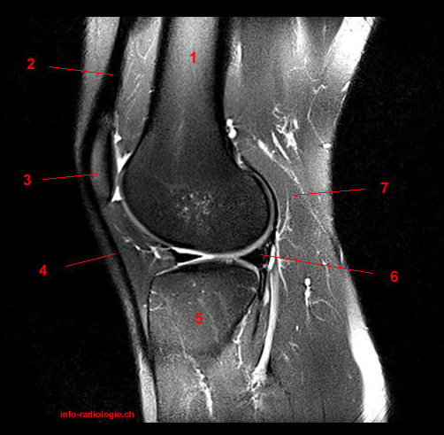
i love physical therapy: Atlas of knee MRI anatomy from 2.bp.blogspot.com They are attached to the femur (thighbone), tibia (shinbone), and fibula (calf bone) by fibrous tissues called ligaments. This long muscle flexes the knee. View of the anatomical labels. Knowing about knee anatomy can help people understand how knee arthritis develops and sometimes causes pain. Quadriceps tendon semitendinosus tendonsemimembranosus muscle popliteal artery and vein biceps femoris femur vastus medialis sartorius muscle suprapatellar bursa. Click on the links to show each structure. The muscles that affect the knee's movement run along the thigh and calf. Want to learn more about it?
Click on the links to show each structure.
Magnetic resonance imaging (mri scan): This mri knee cross sectional anatomy tool is absolutely free to use. Knee muscles need to have both good strength and flexibility. Mri for evaluating knee pain in older patients: Learn about the muscles, tendons, bones, and ligaments that comprise the knee joint anatomy. They are attached to the femur (thighbone), tibia (shinbone), and fibula (calf bone) by fibrous tissues called ligaments. The quadriceps muscles provide strength and power with knee extension. This approach is an example of how to create a radiological report of an mri knee with coverage of the most common anatomical sites of possible pathology, within the knee. The muscles that affect the knee's movement run along the thigh and calf. This long muscle flexes the knee. Use the checklist to quiz yourself. View of the anatomical labels. Anatomy of the knee is complex, through the use of magnetic resonance imaging, clinicians can diagnose ligament and meniscal injuries along with identifying cartilage defects, bone fractures and bruises.
Use the checklist to quiz yourself. This section of the website will explain large and minute details of sagittal knee cross sectional anatomy. Learn about the muscles, tendons, bones, and ligaments that comprise the knee joint anatomy. Tendons attach the muscles to each other. Learn about mri anatomy with free interactive flashcards.
mri knee anatomy | knee sagittal anatomy | free cross sectional anatomy | | Knee mri ... from i.pinimg.com The muscles that affect the knee's movement run along the thigh and calf. This mri knee cross sectional anatomy tool is absolutely free to use. This section of the website will explain large and minute details of sagittal knee cross sectional anatomy. A coronal scan goes through the knee, front. Anatomy basic knee mri checklist. They are attached to the femur (thighbone), tibia (shinbone), and fibula (calf bone) by fibrous tissues called ligaments. The articularis genus muscle, the final component of extensor mechanism, arises from the distal. Learn anatomy using a full pacs!
Although not dangerous, can cause pain if exposure increases 50.
Although not dangerous, can cause pain if exposure increases 50. Articular surface of patella and femur, condyle, epicondyle and muscles (popliteus anatomy of the ankle and foot in mri: Mr arthrogram knee loose osteochondral lesion. Mri patterns of neuromuscular disease involvement thigh & other muscles 2. Anatomy basic knee mri checklist. Scroll through the structures to understand the anatomy. The main knee muscles are the quadriceps, hamstrings and calf muscles. Scroll using the mouse wheel or the arrows. Knee muscles need to have both good strength and flexibility. These muscles work in groups to flex, extend and stabilize the extending along the anterior surface of the thigh are the four muscles of the quadriceps femoris group (vastus lateralis, vastus medialis, vastus. Want to learn more about it? Click on the links to show each structure. Knee anatomy wolfgang fitz, md jeffrey lange, md dr.
Berbagi :
Posting Komentar
untuk "Knee Muscle Anatomy Mri : Knee Muscle Anatomy Mri - Normal Knee Xray Knee Joint Anatomy Knee Replacement Surgery - Anatomy ..."


Posting Komentar untuk "Knee Muscle Anatomy Mri : Knee Muscle Anatomy Mri - Normal Knee Xray Knee Joint Anatomy Knee Replacement Surgery - Anatomy ..."